Traumatologists of Sant Joan de Déu - Barcelona Children’s Hospital reconstruct a girl’s pelvis with a piece of her fibula
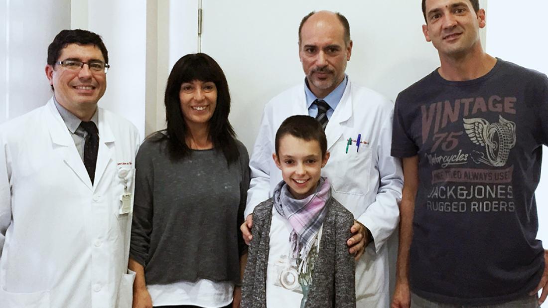
A team of the Orthopaedic Surgery and Traumatology Service of Sant Joan de Déu - Barcelona Children’s Hospital formed by Drs Jorge Knorr, Francisco Soldado, Ferran Torner and Pedro Doménech has reconstructed the pelvis of a girl affected by a tumour, using a piece of the patient’s own fibula.
A team of the Orthopaedic Surgery and Traumatology Service of Sant Joan de Déu - Barcelona Children’s Hospital formed by Drs Jorge Knorr, Francisco Soldado, Ferran Torner and Pedro Doménech has reconstructed the pelvis of a girl affected by a tumour, using a piece of the patient’s own fibula. The operation allowed the surgeons to completely remove the tumour, increasing the chances of a cure in this way and reducing the risk of metastasis. The patient is an 11-year-old girl who was diagnosed with Ewing’s sarcoma, one of the most common bone tumours in adolescents, located in the pelvis.
In these cases, physicians have traditionally ruled out removal of the tumour because there are many nerves, major arteries and viscera in the pelvis area, and also because removal produces a severe alteration of gait. Accordingly, until now it has been chosen to treat patients with radiotherapy. In this case, however, the surgeons decided to remove the tumour and reconstruct the pelvis with a piece from another of the patient’s own bones after meticulously planning the operation with the help of a 3D model of the affected area, which allowed determination of the tumour’s extension and planning of the reconstruction strategy.
The operation took six hours and was carried out in various phases. In the first phase the surgeons removed a piece of the fibula of the leg opposite the area of the pelvis affected by the tumour, for its subsequent transplantation. Then they removed the part of the pelvis affected by the tumour, fixing and ensuring the stability of the bone with a titanium rod. After that they set in place the piece of fibula, folded in half, by levering it in, performing microsurgery to connect it to the blood vessels of the pelvis so that it will grow – an especially important aspect since the patient is a child and is still in the process of development – and so that it will adapt to the loads supported by the pelvis. In this way, the operating team provided a lifelong solution to the patient.
