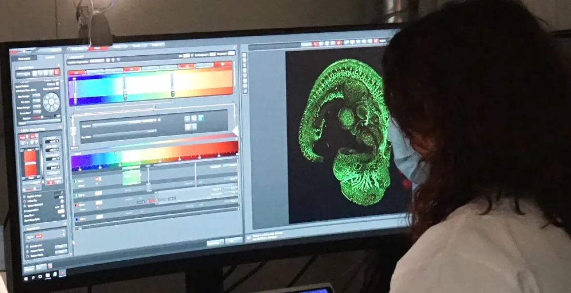
The SJD Barcelona Children’s Hospital’s Confocal Microscopy Unit provides services for researchers in any discipline, including biomedicine, microbiology and even biomaterials, for reasearches that require the use of new technologies in the field of confocal and super-resolution microscopy.
Our team is also available for diagnosis of any disease, helping pathologists and geneticists on the path to diagnosis. This is especially relevant in the field of rare diseases, which are generally complicated to diagnose.
The unit’s facilities are available to researchers from businesses and public and private institutions, including those belonging to the SJD Research Institute and the hospital itself.
Our equipment is state-of-the-art, including advanced optical microscopy systems, sample preparation and maintenance equipment, and image and data processing resources. We also provide confocal nanoscopy, which enables us to substantially improve the resolution of confocal microscopy, allowing individual structures inside cells to be visualised with a resolution of up to 60 nanometres.
We are supported by the hospital’s Anatomical Pathology Department for the sample preparation process and control samples, including cell cultures from the Biobank.
"Our close collaboration with Anatomical Pathology and the hospital’s Biobank makes this an unmatched setting for the study of cell biology and the pathology and aetiopathology of diseases"
The Confocal Microscopy Unit is part of the Genetic and Molecular Medicine Department and the Daniel Bravo Centre for diagnosis and research in minority diseases.
We assist you throughout the process
- We assess you or prepare your samples in the optimum way to ensure you can study them with guarantees.
- Feel free to ask us about the techniques and equipment we use to obtain the best results.
- We will be with you to answer any questions when your samples are under the microscope.
- You have the option of using our equipment unaccompanied in case you demostrate experience in the field.
Technical features
Our staff will help you in the use of our equipment or you can use it independently if you have the necessary knowledge.
Equipment
Our staff will help you in the use of our equipment or you can use it independently if you have the necessary knowledge.
- Confocal laser scanning microscope Leica TCS SP8 with a high speed module and STED 3X (nanoscopy) and HyVolution (super-resolution) modules. Laser lines: 405 nm, 488 nm and a white laser with excitation at 470-670 nm. Depletion lines: 592 nm , 660 nm and 775 nm. Two hybrid detectors.
- Leica DM5500B microscope and DFC7000T colour digital camera for detecting transmitted light, polarised light and fluorescence images.
- Workstations for processing and analysing confocal images (LAS X, Fiji/ImageJ, Huygens).
- Auxiliary equipment: incubator installed on the microscope with temperature and CO2 control, centrifuge, laminar flow cabinet, freezer and CO2 incubators.
Check the various techniques that we can offer you for the study of samples.
Microscopy tecniques
Check the various techniques that we can offer you for the study of samples.
- Multichannel confocal and transmitted light imaging of living cells or fixed samples.
- Three-dimensional analysis and relief maps.
- Multidimensional in vivo experiments: tracking, intracellular calcium, membrane potential, others.
- High-speed confocal microscopy. Protein colocalisation studies.
- Quantitative fluorescence studies and calculation of different parameters.
- F techniques: FRET, FRAP, photoactivation.
- Analysis of the absorption and emission spectra of fluorescent substances.
- Microscopy in multi-position experiments and mosaic acquisition.
- Transmitted light optical microscopy: BF, DIC and polarised light.
- Conventional fluorescence microscopy.
- Image processing and quantification with specialist programs: LAS X, ImageJ, Fiji.
- Nanoscopy and super-resolution studies: HyVolution, STED.
Obtaining good results requires good sample preparation, do not hesitate to contact us.
Sample preparation
Obtaining good results requires good sample preparation, do not hesitate to contact us.
- Preparation of samples of different types of material (biological and non-biological) for optical and fluorescence microscopy.
- Chemical fixation and cutting of biological materials.
- Immunofluorescence in cells and tissues.
- Methodologies for in vivo analysis of cells and physiological studies: specific fluorescent dyes, tagging of ions (calcium), lectins and fluorescent proteins.
Useful documents
We are a Leica Confocal Microscopy training centre.
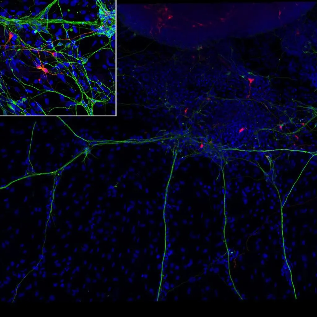
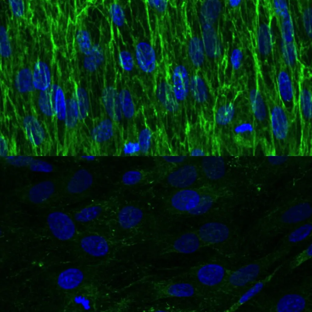
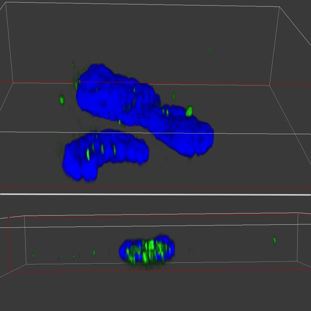
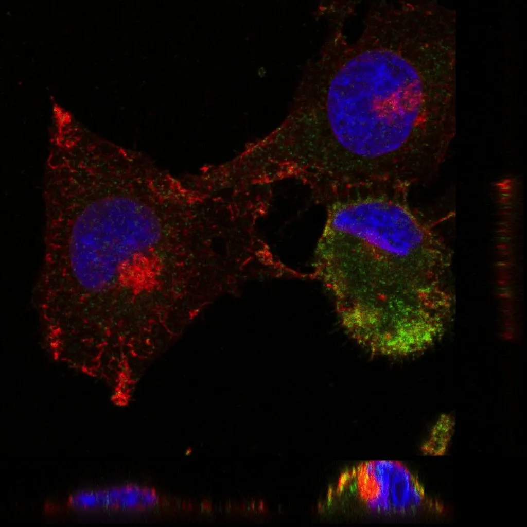
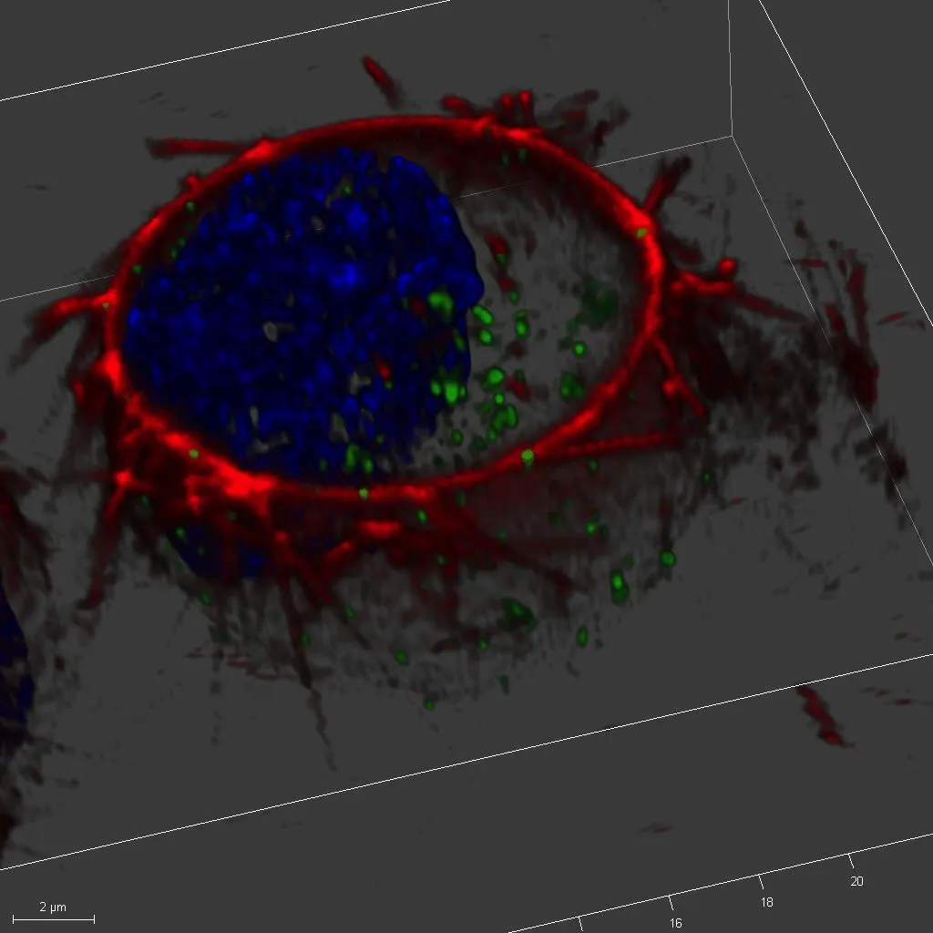
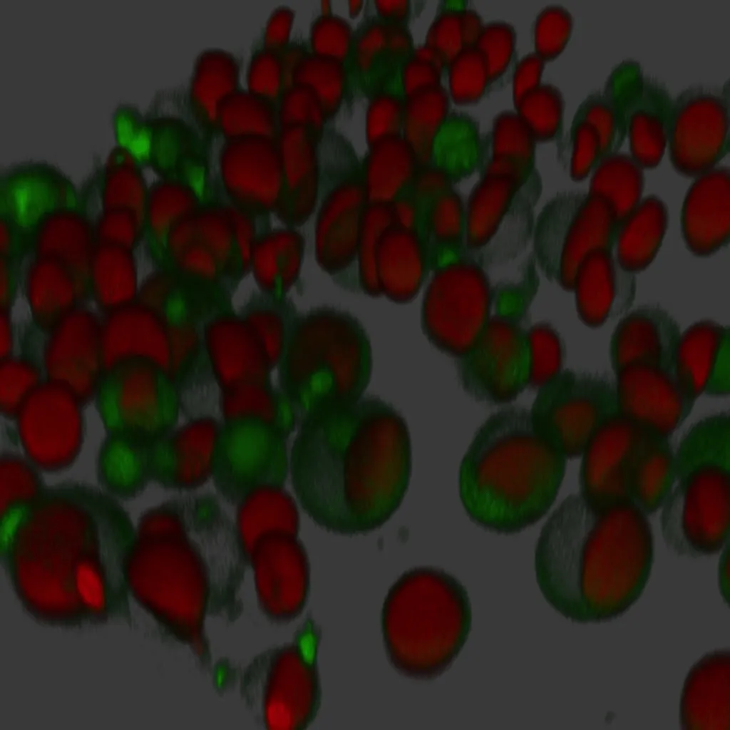
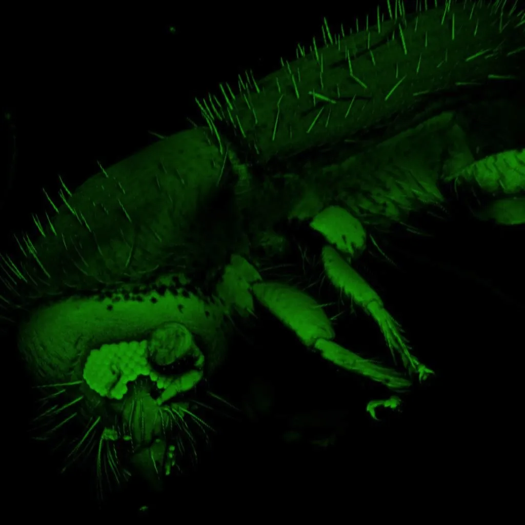
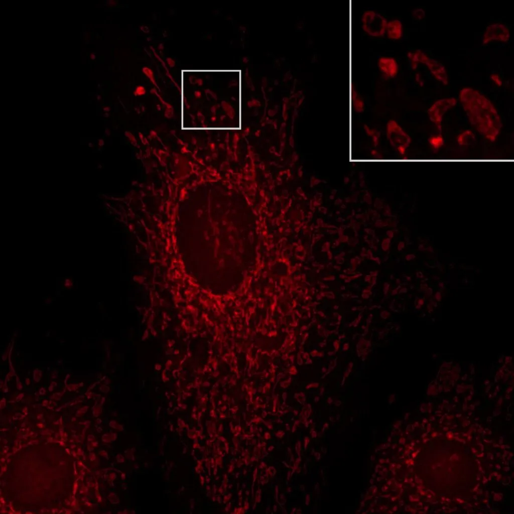
Sample gallery
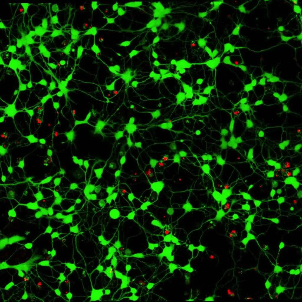
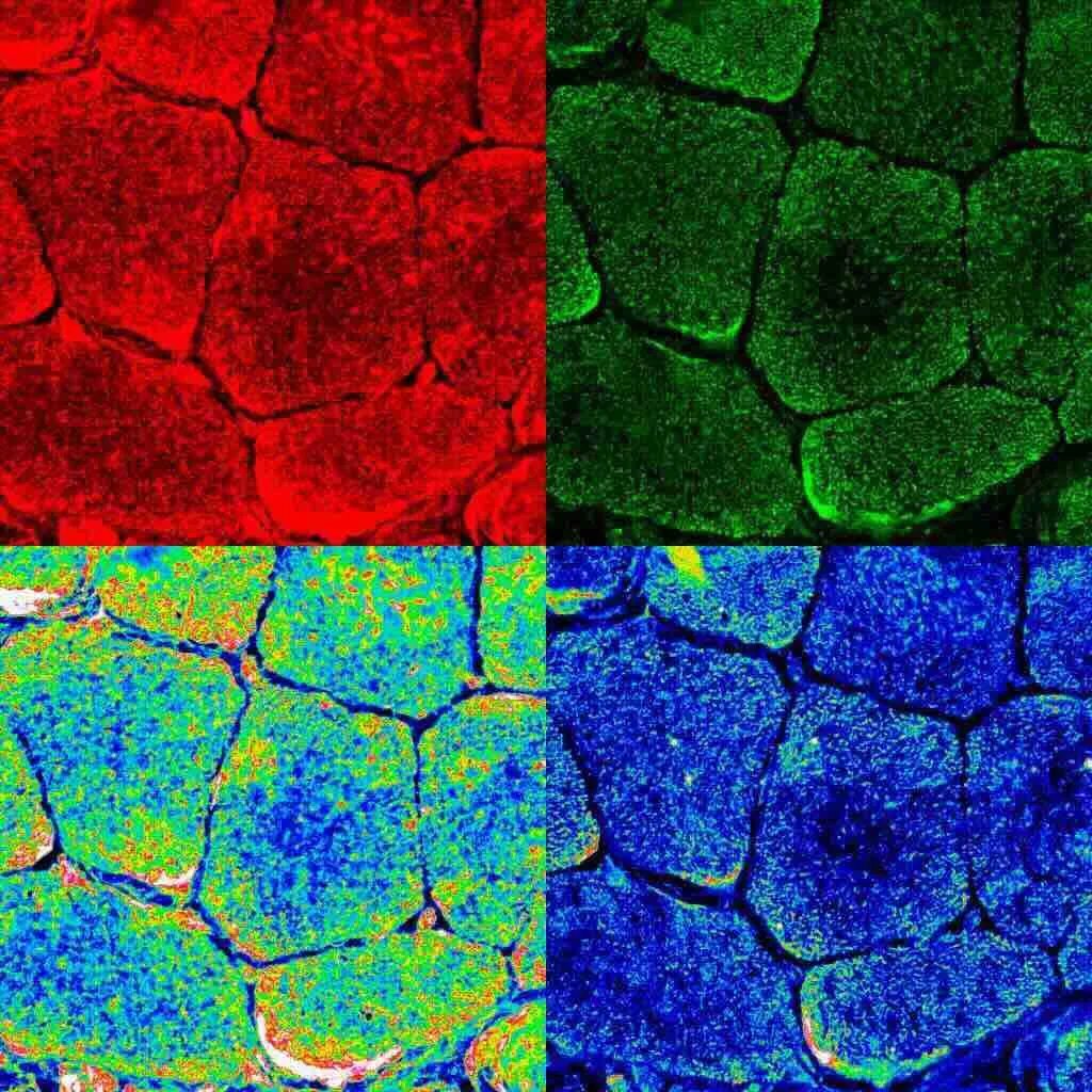

© These images are the property of SJD Barcelona Children’s Hospital, the Sant Joan de Déu Research Foundation or an organisation associated with it, or third parties who have given the Hospital and the Foundation permission to use this content. All rights reserved. The reader may not use them for commercial purposes.
ALBA Synchrotron technology enables the study of subcellular alterations in patients with muscular dystrophy
A research team, made up of researchers from the Neuromuscular Diseases Applied Research Laboratory, the Neurology Service and the Confocal Microscopy and Cell Imaging Unit of the Genetics Service of the Sant Joan de Déu Barcelona Hospital and the Sant Joan de Déu Research Institute, works at the ALBA Synchrotron to study subcellular alterations in patients with muscular dystrophy thanks to its 50nm spatial resolution.
