SJD Barcelona Children’s Hospital, Clínic and UPF create a system that provides more precision, shortens the duration of surgery and reduces the mortality rate
A team of healthcare professionals from BCNatal, a consortium formed by SJD Barcelona Children’s Hospital and Hospital Clínic, has for the first time in the world developed a three-dimensional surgical planning and navigation system for surgery in unborn babies, with the collaboration of BCN-Medtech at Universitat Pompeu Fabra (UPF)
Previously, surgery teams only had ultrasound to guide their entry into the uterus and movement of the surgical instruments towards the foetus. Now, however, the new system allows more precision, shortens the duration of the intervention and, in the future, will improve outcomes by making fetal surgery more accessible.
A GPS that guides you to the point to be operated on quickly and safely
In every pregnant woman, the placenta is located in a different place, her blood vessels are never the same, and the way they connect to the foetus and umbilical cord also varies from pregnancy to pregnancy. The foetus will also always be in a different position in each case, and floating in the amniotic fluid. The foetus is surrounded by very delicate membranes, which can only be pierced once in order not to risk a miscarriage.
Therefore, when a foetus has a life-threatening disease and requires urgent intervention in the mother’s womb, the fetal surgeon faces a great challenge. The surgeon has to pinpoint very accurately where to enter the uterus and, once inside, they have few reference points to help them move around safely.
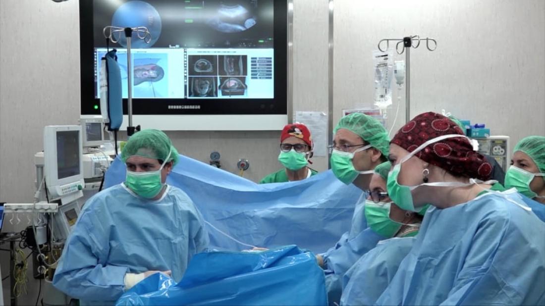
This new system provides a virtual reconstruction of the pregnant woman’s placenta from an MRI scan and an ultrasound. With this 3D map of the placenta, before the intervention, the fetal surgeon has a much more accurate view of the position of the placenta and the umbilical cord, and so is able to analyse the best point of entry to the placenta for access the foetus.
Once in the operating theatre, the surgeon uses a three-millimetre endoscope, specially designed for fetal surgery. The field of vision of fetal endoscopes is very limited and continuous guidance on their position inside the uterus is therefore necessary. This was previously done with ultrasound, but the new system allows for additional 3D navigation which provides information that was not available before. This is because the endoscope has a built-in sensor at the end which is detected by external antennas. Based on complex algorithms and mathematical formulas, this makes it possible to synchronise the virtual reconstruction of the placenta with the actual movements of the surgical instruments.
This is a major breakthrough and a first in the world in fetal surgery, and illustrates how new technologies are essential to achieve safer surgery with systems that provide the surgeon with constant assistance in order to greatly reduce risks.
Twin-to-twin transfusion syndrome
The new navigator is particularly useful for extremely complex fetal surgery interventions, such as to correct twin-to-twin transfusion syndrome, which occurs in 10-15% of pregnancies of monochorionic twins (those who share the placenta). In this case, one of the foetuses constantly passes blood to the other, which almost always causes the death of the twins. With fetal surgery, the situation can be reversed and the survival of at least one of the two foetuses can be achieved in more than 90% of cases.
“The new surgical GPS helps guide surgeons in the operating theatre and also helps them to identify the best entry point, the exact location of the start of the umbilical cords and the blood vessels of the placenta, so the surgery can be completed more easily and with a greater chance of success”, explains Miguel Ángel González Ballester, researcher at ICREA and project coordinator at UPF.
So far in 2019, BCNatal has carried out a total of 20 fetal surgeries with this new system. The results of the technological innovations have been accepted in various international scientific journals on bioengineering. The clinical innovation was presented at the fetal medicine world congress in June of this year and the first series is being prepared for publication.
According to Eduard Gratacós, director of BCNatal, "This is one of the research projects for technological innovations we have been developing for the last four years, thanks to funding from the Fundación Privada Cellex, which are leading to a new generation of techniques that will revolutionise the current way of performing fetal surgery, and very likely other very high-precision surgical interventions which still greatly depend on the personal skill of the surgeon".
BCNatal, formed by SJD Barcelona Children’s Hospital and Clínic, is a pioneering centre in fetal surgery research and clinical practice. BCNatal has developed and performed several of the current techniques for the first time in the world. It hosts doctors from all five continents for specialised medical training, and performs more than 100 fetal surgery interventions a year on national and international patients, with a total track record of 2,000 such interventions.
Related news


