The SJD Barcelona Children's Hospital stops a young girl's blindness from progressing further using a nerve graft taken from her leg
For the first time ever in Spain, surgeons treating a pediatric patient used the sural nerve graft technique to connect a nerve behind the ear with the cornea, thereby recovering corneal sensation.
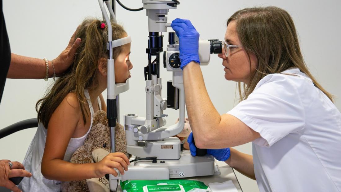
Staff at the SJD Barcelona Children's Hospital have used a groundbreaking technique on a young girl from Valls (Tarragona) who, due to hypoplasia or a poorly developed trigeminal nerve (involved in corneal sensation), was going blind. They managed to stop the vision loss in its tracks, even improving the girl's sight by 10%. The team of surgeons and ophthalmologists took a segment of the girl's sural nerve (located in the leg) and spliced it together with a nerve located in the ear (the great auricular nerve) and with the cornea to reestablish corneal sensation.
Sensation in the cornea is a key aspect of eye health, as it activates the blink reflex to protect against foreign bodies and to stimulate tear production. As she has no corneal sensation, six-year-old Elsa is unable to blink instinctively. This frequently causes keratitis, or ulceration, which further damages the cornea every time it occurs. For Elsa, this has meant significant loss of vision. In 2022, she had only 10% of her vision. To prevent and offset these lesions, Elsa had to use protective eyewear, known as moisture chamber glasses, as well as artificial tears and ointments every hour.

Corneal neurotisation
In cases of hypoplasia, the indicated treatment is corneal neurotisation. This procedure consists of taking a healthy nerve and redirecting it to the cornea to assume the role of the damaged nerve and restore proper function.
‘Traditionally, in classic neurotisations,’ explains Ophthalmologist Ester Casas, ‘the sural nerve is used to connect the facial nerve that is closest to the eye to the cornea, such as the supraorbital or supratrochlear nerve. In Elsa’s case, however, this procedure was not indicated, as these two nerves were also affected by hypoplasia.’ Professionals therefore had to connect the sural nerve to a sensory nerve that was further away, located behind the right ear (the great auricular nerve), and then connect that to the cornea on the same side. This was the first time in Spain that the procedure had been done on a child. ‘To perform this procedure, we had to use microsurgical techniques which, in pediatric cases, is always a challenge due to the smaller size of all of the involved tissues,’ adds Marisa Manzano, Plastic Surgeon in the Pediatric Surgery Department at the SJD Barcelona Children's Hospital.
The operation took place on 18 January 2024, lasting a total of six hours and involving three ophthalmologists and two plastic surgeons. ‘If the procedure is a success, the patient normally takes some time to regain sensation in the cornea. In Elsa's case, her mother noticed signs of sensation after just four months, which was confirmed after a check-up appointment,’ notes Casas. ‘A child’s neural plasticity favours and aids faster recovery than in adults,’ adds Manzano.
The girl's corneal sensation has been recovered, she develops much fewer lesions, and her vision has improved. Now she can be more independent in her activities of daily life without the need to always wear eye protection.
Related news


