Surgeons at SJD Barcelona Children’s Hospital remove an immature undeveloped fetus from a baby, in a complex case
The patient with this unusual disorder was diagnosed thanks to the Fetal Medicine team at SJD Barcelona Children’s Hospital. In the ultrasound at 20 weeks, which reveals many anatomical features of the fetus in detail, a lump was detected that initially looked like a tumour. The child’s development was closely monitored during pregnancy and his family received support.
Initially the aim was to define the type of malformation or tumour in terms of size and characteristics and the impact its growth could have on the development of the fetus. During prenatal development, it was found not to be affecting the fetus and there were no complications at either birth or in the postnatal stage.
After the birth of the child, various diagnostic imaging tests were performed to determine the final diagnosis. The medical team suspected a mature teratoma (a non- malignant tumour) and also a possible case of fetus in fetu, i.e. that the undeveloped fetus of a twin had been “absorbed” by the brother during the pregnancy.
After carrying out the specific studies, the second hypothesis was confirmed and work began with the 3D Unit, as the lump, located in the abdominal area, was close to the baby’s vital organs and it was prudent to plan the surgical intervention.
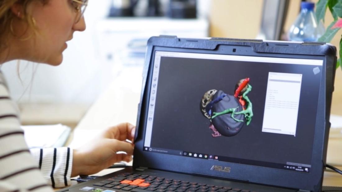
A prior study and the 3D printed model, keys to planning the surgery
After birth, the baby made good progress. As there was no risk to the baby’s health and having established that the lump was not growing, the decision was made to operate after nine months, in order to reduce the risks in the application of anesthesia and for the patient’s recovery.
During that time the location of the undeveloped fetus was studied. In this case, it was treated with the same consideration as a tumour, as it had to be removed, both to prevent it from causing pressure in the child’s abdomen and due to the risk of it beginning to grow at some point.
The 3D Unit’s engineering and planning, Diagnostic Imaging, Fetal and Neonatal Surgery and Oncology Surgery teams all worked together on the pre-operative study.
The final magnetic resonance imaging provided accurate views of the viscera displaced by the undeveloped fetus and also located the aortic artery, the vena cava, the liver and the kidneys. The case was also studied by angiographic techniques to determine how the blood supply arrived at the lump, as it was vascularised. It was confirmed that there were no large arteries connected to the lesion and surgery was scheduled.
The main challenge was to remove the lump while avoiding bleeding of any kind, and the intervention was successful.
3D: a tool to reduce both surgery and patient recovery times
Two days after the intervention, the child was able to eat normally and the parents said he was less restless and uncomfortable.
The planning meant that the surgical team knew what to expect before the intervention, helping to avoid unforeseen situations. In addition, the surgery was relatively brief and the child’s recovery quick and successful. Not all cases require a pre-operative analysis, but in this case, obtaining previous volumes and images provided information which facilitated the work of the medical team.
“In many cases the planning is done virtually, but this time a 3D print-out was made of the object. With these techniques we can make lots of calculations and in the case of a lump or a tumour, one of the most important things we need to know is the volume of tumour to be removed”, explains the Head of Research and Innovation in Diagnostic Imaging, Josep Munuera.
According to Munuera, “The impact of these techniques is that they save surgery time and also save time in the children’s subsequent recovery. So far we have worked with oncology, maxillofacial and trauma surgery and neurosurgery, but in the next few years the number of indications and applications of 3D techniques will increase”.
Related news


